
Dr. Robert Snyder is a podiatrist with over 30 years of experience; his practice is limited to wound management and limb preservation. He is Professor and Director of Clinical Research and Fellowship Director at Barry University SPM. Dr. Snyder is certified in foot and ankle surgery by the American Board of Podiatric Surgery and is also a board certified wound specialist. Dr. Snyder is immediate past-president of the Association for the Advancement of Wound Care and past-president of the American Board of Wound Management. In addition to his doctorate, he holds an MSc in Wound Healing and Tissue Repair from Cardiff University. Dr. Snyder has published several book chapters over 125 papers in peer reviewed and trade journals on wound care and has been a principal investigator on more than 30 randomized controlled trials.
Snyder_Current Dialogues in Wound Management_2015_Volume 1_Issue 3
PATHOPHYSIOLOGY OF CHRONICVENOUS INSUFFICIENCY
Ulcerations caused by chronic venous insufficiency represent the most common wound type in the lower extremities, accounting for more than two-thirds of all ulcerative conditions. Predisposing factors include a history of deep vein thrombosis, obesity, heredity, and pregnancy, among others.
In venous insufficiency, one-way valves function abnormally, thus creating reflux. The backed-up fluid ultimately leaks out of the vasculature into the interstitial tissues via diapedesis, creating venous hypertension. The resulting edema is the most common finding in this condition. Secondarily, abnormalities of the calf muscle pump, sometimes called the second heart, can also lead to increased venous pressure. Conditions, such as equinus deformity of the ankle, may predispose to calf muscle pump dysfunction.
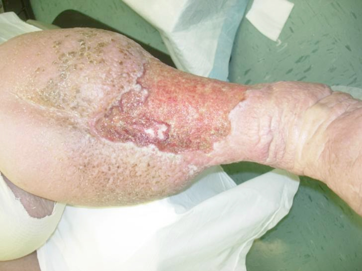
Ulcerations secondary to chronic venous insufficiency normally occur in the gaiter area of the leg, the medial, anterior, and lateral lower leg. The ulcers themselves are usually irregularly shaped, shallow, and in many cases filled predominately with a combination of fibrin and granulation tissue (Figure 1). The wounds may also be extremely painful, especially on ambulation.
Biochemically, it is believed that there is excessive formation of periwound fibrinogen. Although controversial, the fibrin cuff theory holds that fibrin cuffs form around the local microvasculature, blocking penetration of white blood cells and resulting in excessive production of tumor necrosis factor-α, which may predispose to ulcer formation. Pentoxifylline may be used adjunctively, as it has fibrinolytic effects.
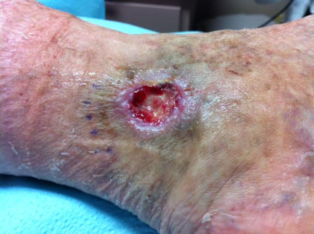 Figure 1. Typical venous leg ulcer in the gaiter area. Note the hemisiderosis and atrophie blanche.
DIAGNOSIS OF VENOUS LEGULCERS
Ulcerations with necrosis, bone, tendon, or muscle usually represent another etiology, such as arterial insufficiency or mixed venous and arterial disease; malignancy; or inflammatory pathology, including pyoderma gangrenosum among others (Figure 2). In these cases, a complete history and physical examination, vascular workup, and wound biopsy are critical.
The extremity itself often resembles an inverted champagne bottle. Scarified or indurated tissue is often observed. This is predominantly an abnormality of the subcutaneous tissue and is referred to as lipodermatosclerosis (Figure 3).
The skin surrounding the ulceration may have a pearly white appearance; this represents a vasculopathy consistent with obstruction of the dermal vessels, referred to as atrophie blanche, which is usually asymptomatic. Hemosiderosis, another common finding, is thought to be caused by leakage of iron from hemoglobin molecules staining the skin. Diagnosis is usually made clinically, but should be substantiated with a venous duplex scan.
TREATMENT OF VENOUS LEGULCERS
The hallmark of therapy for chronic venous insufficiency with ulceration remains compression. A short-stretch bandaging system, such as an Unna boot, a multilayered compression wrap, is recommended during ulcer treatment. Patients often transition to support stockings or garments once the wounds have healed, although adherence may be an issue. Before recommending therapeutic compression, care must be taken to ensure patients do not have an arterial vascular component to their disease or congestive heart failure. An ankle-brachial index (ABI) >.8 mm Hg is an appropriate cut-off value; ABIs of .6–.7 mm Hg may also be considered with less formidable pressures. If the clinician has any concerns about using compression, a vascular or cardiology consultation is recommended. The underlying pathophysiology can be addressed with surgical procedures, such as radiofrequency ablation, among others.
LOCAL WOUND CARE FORVENOUS LEG ULCERS
Many appropriate treatments are available to address the local wound environment. These include standard-of-care therapeutics, such as moist wound healing; antiseptics, such as silver; foams; alginates; negative pressure wound therapy; human skin equivalents and matrices; and dressings that effectively lower excessive protease activity. However, wound bed preparation remains the centerpiece of treatment. This simple but powerful algorithm encapsulates a holistic approach to healing including identifying underlying etiologies, addressing patient-centered concerns, and treating the wound systematically with debridement, control of inflammation and infection, maintenance of moisture balance, and wound edge preparation.
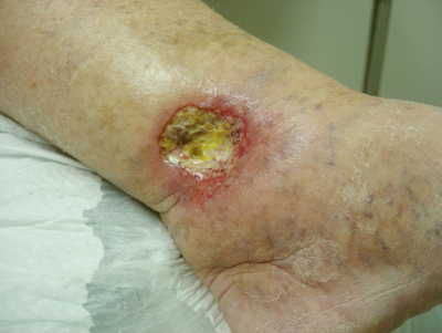
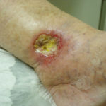 Figure 2. Although the ulcer is located in the gaiter area with evidence of venous insufficiency, the diagnosis in this case is pyoderma gangrenosum.
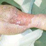 Figure 3. Large venous leg ulcer. Note the inverted champagne bottle deformity of the leg and lipodermatosclerosis.
References
1.Barwell JR, Davies CE, Deacon J, et al. Comparison of surgery and compression with compression alone in chronic venous ulceration (ESCHAR study): randomised controlled trial. Lancet. 2004;363(9424):1854-9.
2.Bergan JJ, Schmid-Schonbein GW, Smith PD, et al. Chronic venous disease. N Engl J Med. 2006;355(5):488-98.
3.Jull AB, Arroll B, Parag V, et al. Pentoxifylline for treating venous leg ulcers. Cochrane Database Syst Rev. 2012 Dec 12;12:CD001733.
4.Schultz GS, Sibbald RG, Falanga V, et al. Wound bed preparation: a systematic approach to wound management. Wound Repair Regen. 2003;11(Suppl 1):S1-28.
5.Snyder RJ. Venous leg ulcers in the elderly patient: associated stress, social support, and coping. Ostomy Wound Manage. 2006;52(9):58-66, 68.
6.Spentzouris G, Labropoulos N. The evaluation of lower-extremity ulcers. Semin Intervent Radiol 2009;26(4):286-95.
7.Tang JC, Marston WA, Kirsner RS. Wound Healing Society (WHS) venous ulcer treatment guidelines: what’s new in five years. Wound Repair Regen. 2012;20(5):619-37.